What is Chronic Venous Insufficiency (CVI)?
Venous insufficiency is typically classified into two types, primary or essential, secondary and post-deep venous thrombosis.
Primary or essential venous insufficiency. Are characterized by the presence of structural changes in the superficial veins of the lower extremities, as expansion and enlargements caused by loss of elasticity and atrophy or disappearance of the valves, the varices are a major element of the vascular disease due to their frequency and the importance of the complications that can result.
It is thought that 15% of the general population suffers from primary venous insufficiency not yet documented the overall percentage for the population with secondary venous insufficiency (post-thrombotic syndrome) following the difficulty of estimating many cases are not documented.
The potential severity of venous insufficiency lies in the complications that can result, such as dermatitis, ulcers, chronic linfeflebedema, repeat thrombosis, and infections of the skin and subcutaneous tissue as common in patients with this disease. CVI is often underdiagnosed.
Anatomy
The venous drainage of the lower limbs is performed by two separate collecting systems, a superficial and a deep, separated by fascia and related by communicating vessels. Venous systems are classified into three groups:
1. Surface or saphenous veins: internal or external magna or less.
2. Deep veins: tibial, peroneal, popliteal, superficial femoral, deep and common.
3. Perforating veins that carry blood from the superficial to the deep system, through the deep fascia.
The superficial veins drain off only 10 to 15% of the blood flow from supra-aponeurotic tissues.
The deep veins of the lower limbs are mostly included in the muscles and follow exactly the path taken named arteries. Both vessels have a connective tissue sheath aponeurotic common drain them of 80 to 85% of the venous blood. At the level of the legs there are two veins for each artery, the common femoral vein at the level of Scarpa's triangle receives the deep femoral and saphenous to iliac become as it passes through the inguinal ligament, the union of both iliac form the inferior vena cava in the right atrium ends.
Iliocaval sector may have one to three valves or lack of them, which supports the importance of the cardiothoracic aspiration or any increase in abdominal pressure hemodynamics. The system-lumbar vertebre plays a major role in the return to the inferior vena cava, which is increased with low obstructions.
Perforating veins. They have one to four valves directed toward the deep venous system. In the legs there are 16 constant perforating veins, which may become insufficient.
Valve apparatus
What predominantly characterizes veins is its valve apparatus. Venous valves are semilunar folds formed by the lining, arranged in pairs facing each other, whose primary mission is to guide the direction of the current vein.
Physiology
Veins of higher circulation ensures four functions:
1. The venous blood return from the distal end toward the right heart capillary.
2. Control extravascular fluid volume, level with exchange capillary venules and maintaining the interstitial fluid reabsorption in the arteriolar filtered.
3. Reservoir function to store and distribute blood mass as the body's needs.
4. Superficial veins play an important role in thermoregulation.
Venous return
Influences centrifuges. Gravity, abdominal pressure, external compression, elasticity, distensibility, collapsibility, course length, bivalence hematopropulsivos mechanisms.
Inadequacies centripetal. Aspiration: cardiopulmonary. Acceleration: venomotricidad, arterial pulsations, muscle activity,
Pathophysiology
There is an imbalance between the influencing factors of centripetal and centrifugal influence, especially in orthostatic position and ambulation. The symptoms are due to a reduced demand capacity (kidnapping with periodic demand deficit).
The main cause of chronic venous insufficiency venous stasis is due in turn to valve damage, either secondary to varicose ineffectiveness, or valvular destruction brought about by a venous thrombosis.
The destruction of the valves produces incompetence of the deep veins and perforating, also causing the normal blood flow of the superficial veins to the deep changes to a reverse abnormally. It is then a superficial venous hypertension, which originates at distal venous stasis, which triggers a series of events anatomical, chemical, mechanical, and blood.
Types of varicose veins
Uncomplicated essential varices of the lower limbs, may take different aspects.
a) Telangiectasis stroke and ways
b) leg veins or varicose veins "broom filament"
c) reticular varices
d) truncal varices
e) Veins of congenital malformations. (Klippel and Weber)
Diagnosis is based on a good history and a proper clinical examination.
Symptoms and signs
Functional
Physical
Medical record
Etiological factors
Differential diagnosis of pain. Whether a person with varicose veins have leg pain does not necessarily mean that they are the cause varicose veins. The most common disease that causes pain and usually attributed to ectatic varices is the nerve root compression due to prolapsed intervertebral disc, or a osteoarthritis of the lumbar spine. Other diseases that cause confusion to family physician include gout, flat feet, heel calcáceo, gonarthrosis, alcoholic or diabetic neuropathy, and degenerative musculoskeletal diseases.
Physical Examination
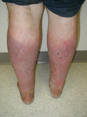
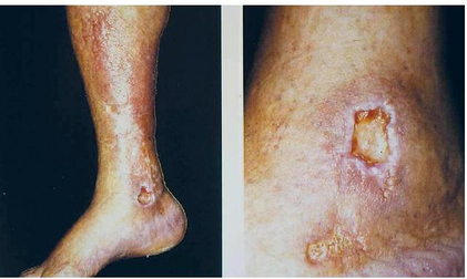
It must be always with the patient standing, observe if morphological changes in the pelvic limbs and appreciate the distribution, shape and color of varicose dilations. Are typical tension, edematous thickening and congestion at the ankle and leg, internal malleolar hyperpigmentation. Dry, scaly skin, sometimes escleosis invades subcutaneous tissue. On this cause skin trauma devitalized any flebostática ulcer, trauma and if the result is about one varicorrhage ectasia.
The examination should be complete, to:
a) valvular insufficiency of the internal and external saphenous. (Test Schwartz)
b) valvular insufficiency of both saphenous arch. (Trendelenburg test)
c) perforator valvular insufficiency. (Trendelenburg test)
d) Permeability of the deep venous system. (Proof of Perthes)
Diagnostic techniques
Treatment Options
Specific Treatment
Doctor: Preventive measures and medications before phlebothropics, oils, diuretics and anti-inflammatory when cases require, whether there will be handled with cures ulcers, colagenizados dressings, etc.. In cases of infections will provide specific antibiotics added. Surgical: Sclerotherapy and surgery.
Bibliography
1. Latorre J. "Anatomy, physiology and pathophysiology of the venous system." Come Insuf Cr lower limb. ed. Uriach Doc Lab Center. Barcelona 1986. 21-59.
2. Bassi G L. Actualité biologique. Les Mécanismes veineux du retour. Path Biol 10, No. 7-8: 695-703
3. Barnes R W. Diagnosis of deep vein thrombosis. Vasc 1980.1:20-24 Diag Ther.
The venous drainage of the lower limbs is performed by two separate collecting systems, a superficial and a deep, separated by fascia and related by communicating vessels. Venous systems are classified into three groups:
1. Surface or saphenous veins: internal or external magna or less.
2. Deep veins: tibial, peroneal, popliteal, superficial femoral, deep and common.
3. Perforating veins that carry blood from the superficial to the deep system, through the deep fascia.
The superficial veins drain off only 10 to 15% of the blood flow from supra-aponeurotic tissues.
The deep veins of the lower limbs are mostly included in the muscles and follow exactly the path taken named arteries. Both vessels have a connective tissue sheath aponeurotic common drain them of 80 to 85% of the venous blood. At the level of the legs there are two veins for each artery, the common femoral vein at the level of Scarpa's triangle receives the deep femoral and saphenous to iliac become as it passes through the inguinal ligament, the union of both iliac form the inferior vena cava in the right atrium ends.
Iliocaval sector may have one to three valves or lack of them, which supports the importance of the cardiothoracic aspiration or any increase in abdominal pressure hemodynamics. The system-lumbar vertebre plays a major role in the return to the inferior vena cava, which is increased with low obstructions.
Perforating veins. They have one to four valves directed toward the deep venous system. In the legs there are 16 constant perforating veins, which may become insufficient.
Valve apparatus
What predominantly characterizes veins is its valve apparatus. Venous valves are semilunar folds formed by the lining, arranged in pairs facing each other, whose primary mission is to guide the direction of the current vein.
Physiology
Veins of higher circulation ensures four functions:
1. The venous blood return from the distal end toward the right heart capillary.
2. Control extravascular fluid volume, level with exchange capillary venules and maintaining the interstitial fluid reabsorption in the arteriolar filtered.
3. Reservoir function to store and distribute blood mass as the body's needs.
4. Superficial veins play an important role in thermoregulation.
Venous return
Influences centrifuges. Gravity, abdominal pressure, external compression, elasticity, distensibility, collapsibility, course length, bivalence hematopropulsivos mechanisms.
Inadequacies centripetal. Aspiration: cardiopulmonary. Acceleration: venomotricidad, arterial pulsations, muscle activity,
Pathophysiology
There is an imbalance between the influencing factors of centripetal and centrifugal influence, especially in orthostatic position and ambulation. The symptoms are due to a reduced demand capacity (kidnapping with periodic demand deficit).
The main cause of chronic venous insufficiency venous stasis is due in turn to valve damage, either secondary to varicose ineffectiveness, or valvular destruction brought about by a venous thrombosis.
The destruction of the valves produces incompetence of the deep veins and perforating, also causing the normal blood flow of the superficial veins to the deep changes to a reverse abnormally. It is then a superficial venous hypertension, which originates at distal venous stasis, which triggers a series of events anatomical, chemical, mechanical, and blood.
Types of varicose veins
Uncomplicated essential varices of the lower limbs, may take different aspects.
a) Telangiectasis stroke and ways
b) leg veins or varicose veins "broom filament"
c) reticular varices
d) truncal varices
e) Veins of congenital malformations. (Klippel and Weber)
Diagnosis is based on a good history and a proper clinical examination.
Symptoms and signs
Functional
- Heaviness and tiredness of legs that increases with standing and heat. Symptoms decrease with cold, lying and walking.
- Hyperesthesia and calf muscle cramps in evening usually due to fatigue.
- Intense itching in the supra-malleolar region that extends half of the leg, and that causes scratching.
Physical
- Varicosities
- Edema initially supramalleolar region of marbling, mainly in the evening, it is necessary to differentiate from edema due to other causes.
- Pigmentation and discoloration of the skin: dermatitis ocher and white atrophy.
- Ulcers supramaleolares especially the medial malleolus with eczematous halo and accompanied by desquamation.
- No temperature increase of the skin, erythema and pain in the ectatic path (varicophlebitis).
Medical record
Etiological factors
- Inheritance varicose n, which is the most important primary cause.
- Difficult trade or profession: work standing, sedentary and heat exposure.
- Date and circumstances of the occurrence. Children (angiodysplasia). After thrombosis (secondary or postflebíticas varices). Pregnancy.
Differential diagnosis of pain. Whether a person with varicose veins have leg pain does not necessarily mean that they are the cause varicose veins. The most common disease that causes pain and usually attributed to ectatic varices is the nerve root compression due to prolapsed intervertebral disc, or a osteoarthritis of the lumbar spine. Other diseases that cause confusion to family physician include gout, flat feet, heel calcáceo, gonarthrosis, alcoholic or diabetic neuropathy, and degenerative musculoskeletal diseases.
Physical Examination
It must be always with the patient standing, observe if morphological changes in the pelvic limbs and appreciate the distribution, shape and color of varicose dilations. Are typical tension, edematous thickening and congestion at the ankle and leg, internal malleolar hyperpigmentation. Dry, scaly skin, sometimes escleosis invades subcutaneous tissue. On this cause skin trauma devitalized any flebostática ulcer, trauma and if the result is about one varicorrhage ectasia.
The examination should be complete, to:
a) valvular insufficiency of the internal and external saphenous. (Test Schwartz)
b) valvular insufficiency of both saphenous arch. (Trendelenburg test)
c) perforator valvular insufficiency. (Trendelenburg test)
d) Permeability of the deep venous system. (Proof of Perthes)
Diagnostic techniques
- No venous Doppler (uroflow study that evaluates speed).
- No venous occlusion plethysmography (blood flow assessment by volumetric alterations registration).
- Venography of lower limbs (radionuclide or radiation).
- Any of the first two must be made first choice always gifted medical centers, given its ease of interpretation, speed and replicability, before performing an invasive study such as venography for the risks involved.
Treatment Options
- Compression Stockings or bandaging
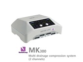
- Home Compression Pump - With Sequential Gradient Inflation Cycles
- Be directed towards prevention, with special hygiene measures vein, such as:
- Maintain body weight within normal limits.
- Do not be too long standing or sitting.
- Do not use belts or tight clothing.
- Constantly lubricate the legs and ankles.
- Raise the footboard of the bed 15 cms.
- Using socks low, medium or high compression, depending on the magnitude of the suffering.
- Perform aerobic exercise frequently (avoid weight lifting).
- Avoid possible to anovulatory intake and hormone supplements.
- No smoking.
- Avoid trauma to legs and feet.
- During the day, elevate the legs 15 cms. every 8 hours, for 10 mins.
- On long trips propelled vehicles, get up and walk for a few minutes every two hours.
Specific Treatment
Doctor: Preventive measures and medications before phlebothropics, oils, diuretics and anti-inflammatory when cases require, whether there will be handled with cures ulcers, colagenizados dressings, etc.. In cases of infections will provide specific antibiotics added. Surgical: Sclerotherapy and surgery.
Bibliography
1. Latorre J. "Anatomy, physiology and pathophysiology of the venous system." Come Insuf Cr lower limb. ed. Uriach Doc Lab Center. Barcelona 1986. 21-59.
2. Bassi G L. Actualité biologique. Les Mécanismes veineux du retour. Path Biol 10, No. 7-8: 695-703
3. Barnes R W. Diagnosis of deep vein thrombosis. Vasc 1980.1:20-24 Diag Ther.
|
|


